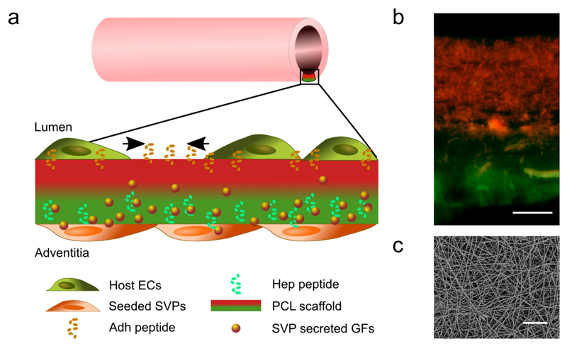
Figure 1. Characterization of the engineered scaffold for blood vessel graft applications.
The bifunctional scaffold is composed of electrospun peptide-conjugated polycaprolactone (PCL) fibres seeded on the abluminal side with patient-derived pro-angiogenic saphenous vein pericytes (SVPs).
As shown in the schematic (a) and the overlaid fluorescence microscopy image (b), the luminal side of the scaffold is mainly decorated with the Adh peptide (in red), to increase host endothelial cell (ECs) adhesion, migration and spreading. The outer layer presents mainly the Hep peptide (in green), which binds and present the SVP-produced growth factors (GFs). SEM image showing the size and distribution of the fibres in the scaffold (top view, c). Scale bars: 50 μm.

