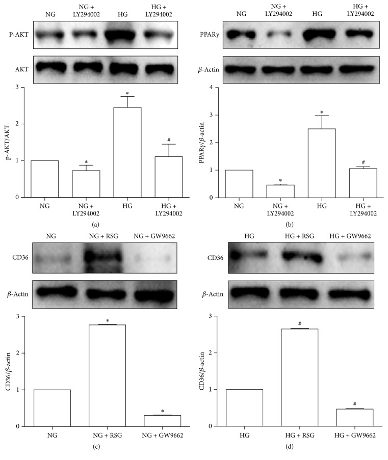
Figure 3.
PPARγ is upregulated by the HG-promoted AKT phosphorylation, which can regulate CD36 expression in HK-2 cells. HK-2 cells were cultured with NG (5.6 mM), NG + LY294002 (15 uM), HG (30 mM), or HG + LY294002 (15 uM) for 48 h. P-AKT, AKT, and PPARγ were examined by western blotting (a, b). HK-2 cells were pretreated with an agonist (RSG, 5 uM)/antagonist (GW9662, 2.5 uM) of PPARγ for 1 h, followed by 48 h of NG (5.6 uM)/HG (30 uM) stimulation for the analysis of CD36 protein levels. Using western blotting, cell lysates were analyzed (c, d). All experiments were repeated thrice. Band intensities were normalized to β-actin band intensity using densitometry. The data were represented as the means ± SD. ∗P < 0.05 versus NG; #P < 0.05 versus HG.

