
Figure 2.
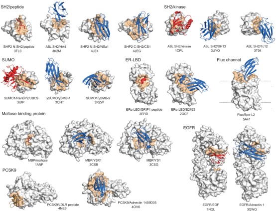
Examples of Monobodies and Adnectins binding to a functional site within the target protein. The target proteins are shown in gray with the epitope in orange. Natural ligands are in red, and Monobodies and Adnectins in blue. The identities of the target molecules and PDB entry codes are indicated. For the Fluc channel structure, the natural ligand, F– ion, is not shown because of its small size.