
Figure 4.
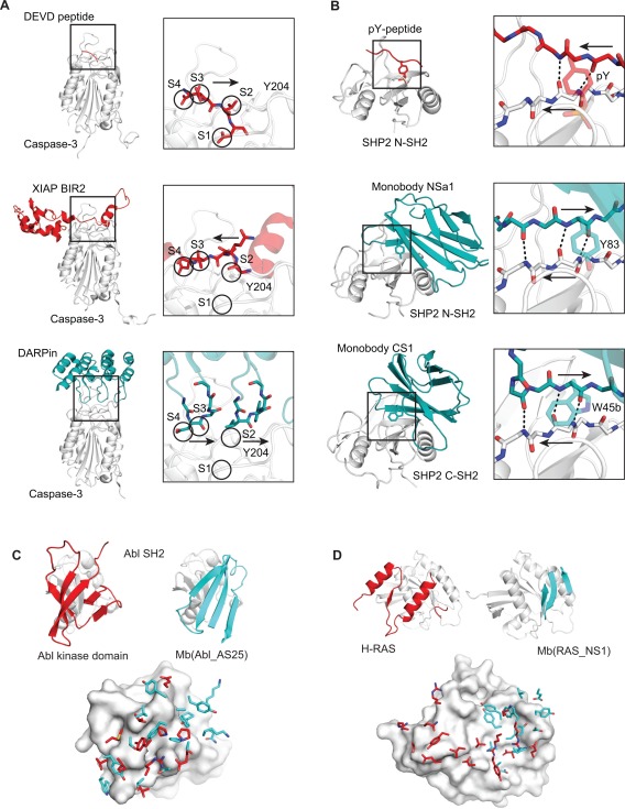
Synthetic binding proteins identify novel modes of protein‐protein interactions. Comparisons of the binding mode of a natural ligand (red) and that of a synthetic binding protein (blue) for Caspase‐3 (A), SHP2 SH2 domain (B), Abl SH2 (C) and H‐RAS (D) are shown. For C and D, the side chains of the ligand and synthetic binding protein located within 4.5 Å of the target protein are shown in the lower panels.