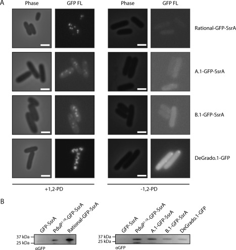
Figure 2.

De novo signal sequences localize GFP to Pdu microcompartments. (A) Phase contrast and fluorescence microscopy of S. enterica expressing Pdu MCPs and signal sequence–GFP fusions as indicated. Scale bars indicate 1 μm. (B) Anti‐GFP western blot of Pdu MCPs purified from S. enterica expressing Pdu MCPs and signal sequence–GFP fusions as indicated.
