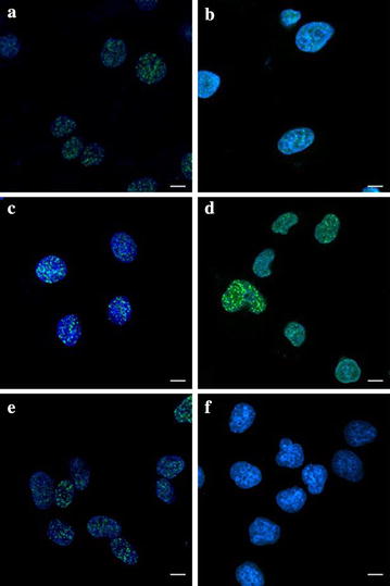
Fig. 2.

Digitized images of γH2AX and pATM foci. After exposure to 2 Gy 12C6+ and incubation 0.5 h for γH2AX and 4 h for pATM, cells were grown and irradiated on cover slips. DNA was stained with DAPI and γH2AX and pATM was detected using an Alexa 488-conjugated secondary antibody after staining using anti-phospho-histone H2AX (Ser-139) and anti-phospho-ATM (ser1981) mAb. a Hela-γH2AX; b Hela-pATM; c HepG2-γH2AX; d HepG2- pATM; e MEC-1-γH2AX; f MEC-1-pATM. Scale bar 15 μm
