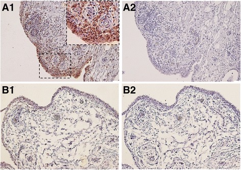
Fig. 1.

S100A11 protein in rheumatoid arthritis (RA) and osteoarthritis (OA) synovial tissue assessed by immunohistochemical (IHC) analysis. Intensive staining for S100A11 was found in the synovial fibroblasts of the lining layer and within the inflammatory cell infiltrates in the RA synovial tissue (A1). The S100A11 expression was negligible or absent in OA synovium (B1). Mouse IgG was used as an isotype control (A2, B2). Representative images of IHC staining are shown at × 200 magnification. For the detailed view the × 400 magnification is shown (n = 6 patients with RA and n = 6 patients with OA)
