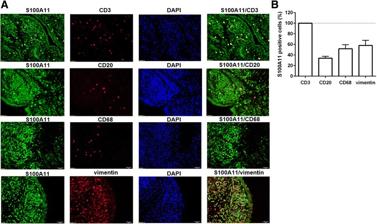
Fig. 2.

Cellular distribution of the S100A11 protein in rheumatoid arthritis synovial tissue. Representative images of immunofluorescence staining (a) for CD68 (macrophages), CD20 (B cells), CD3 (T cells) and vimentin (cells of mesenchymal origin) are shown at × 200 magnification (n = 4). DAPI 4',6-diamidino-2-phenylindole. b Average percentage of S100A11-positive cells presented as mean ± SEM
