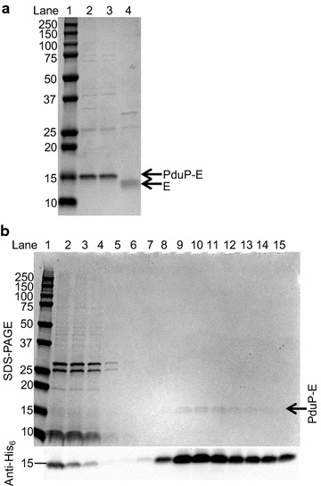
Fig. 6.

Purified PduP-E and non-tagged E from whole cells and purified BMCs. a SDS-PAGE analysis of PduP-E purified from whole cells and non-tagged E after Factor Xa proteolysis. Lane 1 molecular weight markers with sizes of standards in kDa on the left. Lane 2 PduP-E purified from Ec3087 induced with 0.1 mM rhamnose only. Lane 3 PduP-E purified from Ec3087 co-induced with 0.1 mM rhamnose and 0.5 mM IPTG. Lane 4 non-tagged E after Factor Xa proteolysis. Lanes 2–4 were loaded with 1 µg of protein. b SDS-PAGE and anti-His6 Western blot analysis during purification of PduP-E from purified BMCs. Lane 1 molecular weight markers. Lane 2 purified BMCs from Ec3087 co-induced with rhamnose and IPTG (starting material). Lane 3 flow through after binding to the Ni–NTA column. Lanes 4–6 sequential wash fractions containing 20 mM imidazole. Lanes 7–15 sequential elution fractions containing 200 mM imidazole
