
Fig. 1.
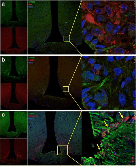
Cellular distribution of GPR120 in the hypothalamus of mice. Tissue sections (5.0 μm) were prepared from the hypothalamic region of lean Swiss mice and were evaluated by indirect immunofluorescence staining using antibodies against GPR120 (a–c, green), NPY (a, red), POMC (b, red), and mannose receptor (c, red). Nuclei were stained with DAPI (blue). In the captions, the arrows indicate cells co-expressing GPR120 and mannose receptor (c). Images are representative of three independent experiments