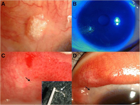
Fig. 2.

a A nodulated mass with calcium depositions was located on superior bulbar conjunctiva. b The cornea was relatively clear with no epithelial defects. c With eversion of the superior conjunctival fornix, a nonabsorbable suture with focal papillary reaction was detected on superior palpebral conjunctiva (black arrow). The suture was removed with slit lamp (white arrow). d After 2 months, the lesion was resolved (black arrow)
