Figure 3.
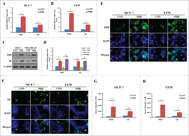
PHB induces AR expression in the ER-positive breast cancer cells. PHB and AR mRNA levels were detected by q-PCR following transfection of plasmid encoding PHB into MCF-7 (A) and T47D (B) cells, and normalized to GAPDH expression. (C, D) PHB protein levels were determined by western blotting following transfection of plasmid encoding PHB into MCF-7 and T47D cells, and normalized to GAPDH expression. Localization and expression of PHB (E) and AR (F) were determined by fluorescence microscopy following the same treatment as (A, B). Nuclei were stained with DAPI. The areas of PHB or AR positive cells in the MCF (G) and T47D (H) cells were analyzed.

