
Fig. 3.
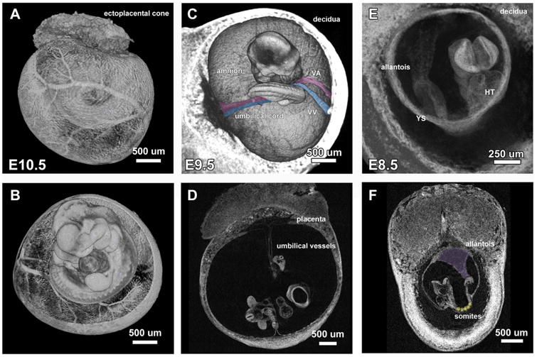
MicroCT imaging of E10.5 to E8.5 mouse embryos within extra-embryonic structures. Iodine contrast E10.5 embryo within yolk sac imaged on microCT revealed (A) the remodeled vasculature on the yolk sac and (B) the orientation of the embryo within the yolk sac. E9.5 embryo imaged within decidua shows (C) the connection of vitelline vein/artery (pseudo-colored in blue/pink) and umbilical vein/artery (pseudo-colored in pink/blue) to yolk sac and placenta and (D) 2D virtual section of the E9.5 embryo within decidua reveals the diameter and length of remodeled umbilical vessels. (E) E8.5 embryo imaged within decidua shows the original orientation of the unturned embryo with the allantois extending from the tail toward the chorion. (F) 2D virtual section of an E8.5 embryo shows the sharp boundary of the somites (pseudo-colored in yellow) and how the allantois extends to the chorion (pseudo-colored in purple).