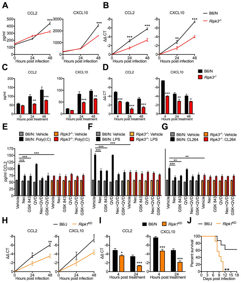
Figure 3. RIPK3-mediated neuronal chemokine expression occurs downstream of multiple TLRs and requires the kinase activity of RIPK1 and RIPK3.
(A–D) CCL2 or CXCL10 expression in B6/N or Ripk3−/− cortical neuron cultures following infection with 0.001 MOI WNV-TX (A–B) or treatment with 1 μg/ml poly(I:C) (C–D), measuring protein in supernatant via Bio-plex multiplex immunoassay assay (A,C) or mRNA expression in cells via qRT-PCR (B,D). N=3–6 replicates/group.
(E–G) CCL2 expression measured by ELISA in cortical neuron culture supernatants after 24h treatment with 1 μg/ml poly(I:C) (E), 1 μg/ml LPS (F), or 1 μg/ml CL264 (G). Prior to addition of TLR agonist, cells were pretreated for 1h with 30 μM Necrostatin-1 (Nec), 100 nM GSK 843, and/or 2 μM QVD. Inhibitors remained in culture medium for the duration of the experiment. As experiments in (C–E) happened concurrently, data with each TLR agonist is compared against a single set of vehicle controls (gray and orange bars), the data for which is repeated in each panel. N=4 replicates/group.
(H–I) CCL2 or CXCL10 expression in B6/J or Ripk1KD/KD cortical neuron cultures following infection with 0.001 MOI WNV-TX (H) or treatment with 1 μg/ml poly(I:C) (I), measured via qRT-PCR. N=4 replicates/group.
(J) Survival analysis in 8 week old Ripk1KD/KD mice and B6/J controls following subcutaneous inoculation with 100pfu WNV-TX. N=7 mice/genotype.
-*p<0.05, **p<0.01, ***p<0.001. Error bars represent SEM. All data are pooled from at least two independent experiments.
See also related figures S4 and S5.

