
Figure 2.
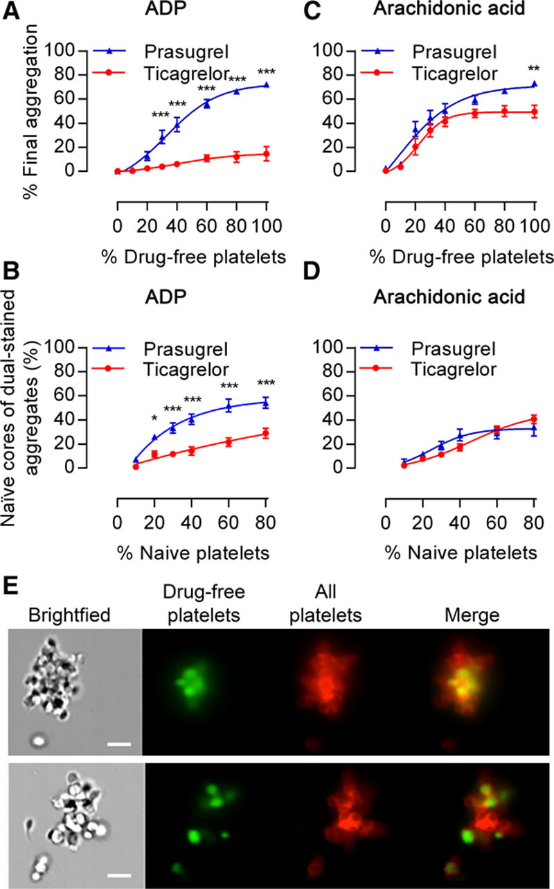
Drug-free platelets restore ex vivo aggregation responses differentially in the presence of prasugrel or ticagrelor. Platelet-rich plasma (PRP) isolated from individuals 6 h after receiving aspirin+prasugrel or aspirin+ticagrelor was mixed with increasing proportions of drug-free platelets and then stimulated. Final aggregation of samples stimulated with (A) ADP (20 μmol/L) or (C) arachidonic acid (1 mmol/L). B and D, Aggregates (%) where cores comprise naive platelets were blind scored and calculated from flow cytometric images of platelet aggregates from corresponding samples. One hundred four aggregates assessed per individual sample, with data presented as mean±SEM and compared by 2-way ANOVA (n=10 samples; *P<0.05, **P<0.01, ***P<0.001). E, Representative flow cytometric imaging (×60 objective) of aggregates formed in response to ADP (20 μmol/L) from 80%:20% mixtures of aspirin+prasugrel–inhibited platelet-rich plasma (PRP) or aspirin+ticagrelor-inhibited PRP obtained 6 h after the last drug dose was administered (red) and drug-free platelets (green). Scale bars, 7 μm.