
Figure 3.
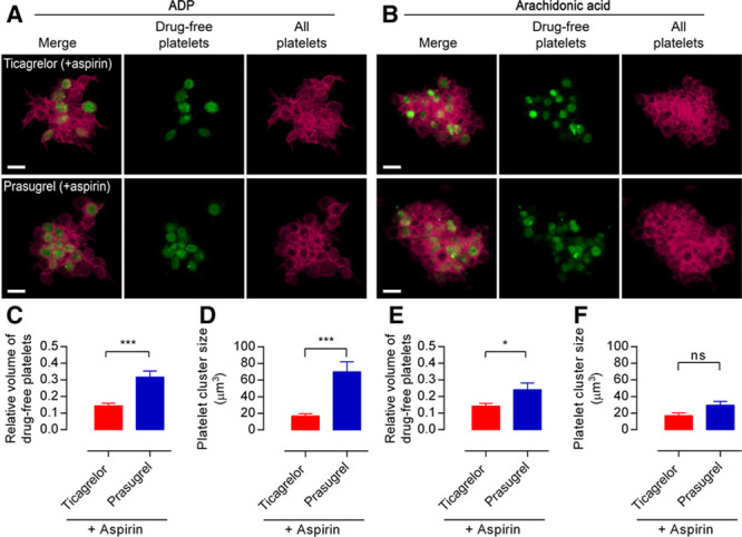
Drug-free platelets form cores within aggregates in the presence of prasugrel but not of ticagrelor. Representative confocal images of (A) ADP-stimulated or (B) arachidonic acid–stimulated aggregates formed from 80%:20% mixtures of aspirin+prasugrel–inhibited platelet-rich plasma (PRP) or aspirin+ticagrelor–inhibited PRP obtained 6 h after the last drug dose was administered (red) and drug-free platelets (green), conditions as in Figure 2. Images were analyzed for (C and E) volume of the drug-free platelet particles relative to the total aggregate volume and (D and F) average size of drug-free platelet clusters. Scale bars, 5 μm. Data are presented as mean±SEM and compared by t test (n=7–10; *P<0.05, ***P<0.001).