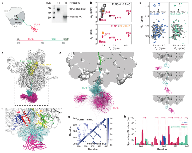
Figure 1. Structural ensemble of a ribosome-bound nascent chain.
(a) Schematic of the FLN5+110 RNC used for the ensemble calculations. The FLN5 sequence is tethered to the ribosome by a C-terminally truncated FLN6751-839 sequence and stalled using the SecM translational-arrest motif27,29. Anti-His western blots of purified FLN5+110 RNC, ribosome-attached with bound prolyl P-site tRNA, and also in its released form. (b) Overlay of 1H-13C correlation spectra of [U-2H; Ileδ1-13CH3 labeled] FLN5+110 RNC (black) with isolated, natively folded FLN5 (pink) and isolated unfolded FLN5∆16 (orange). (c) Overlay of 1H-15N correlation spectra of U-15N-labelled FLN5+110 RNC with isolated FLN5∆12 (blue), and unfolded FLN5-6∆18 (green). (d) NMR chemical shift restrained structural ensemble of FLN5+110 RNC, showing the disordered FLN6 linker (cyan) and the native fold acquired by FLN5 (pink). (Accession codes: PDB ID 2N62; BMRB ID 25748). (e) Close-up view of the ribosomal exit tunnel, highlighting three representative conformations of the nascent chain ensemble (left); the three representative conformations are also shown separately (right). (f) Transient interactions made between the disordered FLN6 linker in close proximity with the ribosomal proteins at the surface. (g) Probability of the formation of inter-residue contacts in the FLN5+110 RNC (shown above diagonal) and in the native state of full length (FL), isolated FLN5-6 (below diagonal) (h) Secondary structure populations of the RNC depicting β-strands (red), α-helices (blue) and polyproline II regions (green); native β-strands are indicated (red arrows).

