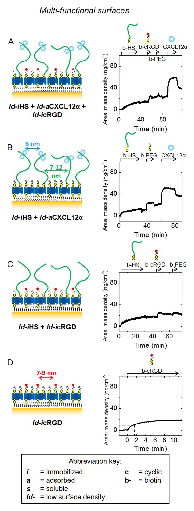
Figure 4. Design and preparation of multifunctional biomimetic surfaces presenting GAG-bound chemokine and integrin ligands.
Schematic presentation of model surfaces (left) used to study the joint effect of HS-bound CXCL12α (ld-iHS + ld-aCXCL12α) and the immobilized integrin ligand cyclic arginylglycylaspartic acid (ld-icRGD) on myoblast adhesion and motility; surface functionalization was followed by SE to quantify areal mass densities (right; see also Table 2). Schemes and SE data are displayed analogous to Fig. 1B. To accomodate all functional molecules, these were presented at moderately lower densities (ld) compared to Fig. 1B; next to surfaces displaying ld-iHS, ld-aCXCL12α and ld-icRGD (A), controls displaying only one or two of the three components (B-D) at comparable surface densities were also prepared. cRGD was immobilized through a PEG-linked biotin; biotinylated polyethylene glycol (b-PEG) was used to back-fill the remaining free biotin-binding pockets on the breadboard.

