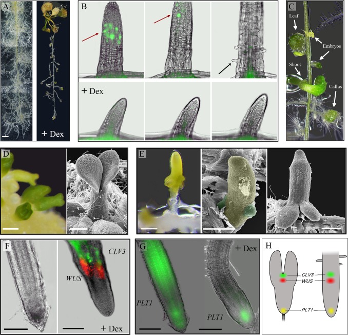
Fig 1. WUS inhibits Arabidopsis root responses to auxin, suppresses cell division and represses the expression of the PLETHORA1 gene, thereby promotes shoot fate.
35S::WUS-GR Arabidopsis seedlings (11- day old) were transferred to a medium supplemented with auxin (0.5μM NAA) in the presence or absence of 10μM Dex. A. Auxin induced massive lateral root formation (left). Activation of WUS by Dex led to inhibition of root formation and root growth (right). Images were taken after 13 days of culturing; separate images were merged. Bar = 2 mm. B. Induction of WUS by Dex suppresses cell division and elongation. Cell-division events in lateral roots were monitored by the cell-cycle marker CYCB1;1::GFP upon culturing on auxin in the presence or absence of 10μM Dex. Images were captured at 24h, 26h and 40 min, and 41h of incubation. Red arrow: dividing cells, black arrow: root hair. Bars = 100 μm (see also time-lapse S1 and S2 Movies). C. WUS in the presence of auxin induces the formation of callus, embryos and shoots. Image was taken after 12 days of culturing on auxin and Dex. Bar = 0.5 mm. D. Somatic embryos formed on wild-type callus exhibit typical basal-apical polarity: cotyledons distal and embryonic radicle proximal to the callus. Bar = 100 μm. E. WUS induces somatic embryo formation with atypical orientation. The embryonic radicle coincided with the lateral-root tip and the cotyledons developed shootward. Scanning electron microscopy views demonstrate the initiation of two cotyledon primordia on the lateral root (root and cotyledon primordia are false-colored yellow and green, respectively). Bars = 200 m. F. WUS activation induces the establishment of an organizing center and stem-cell population. The expression of WUS and CLV3 was monitored by using the 35S::WUS-GR x WUS::DsRed x CLV3::GFP marker line. No signal was detected in the presence of auxin after 12 days of culturing (left). Intense WUS (DsRed) and CLV3 (GFP) signals were detected in the presence of auxin and Dex after 12 days of culturing (right). Bars = 100 μm. G. WUS induction represses the PLT1 expression. 35S::WUS-GR x PLT1::YFP roots incubated for 104h on auxin without (left) or with (right) Dex. On the auxin-only medium, intense signals were detected in the root tip and elongation zone. In the presence of auxin and Dex, the signal was confined to the root tip. Bars = 100 μm. H. Scheme of WUS (red), CLV3 (green) and PLT1 (yellow) patterns of expression in a torpedo-stage zygote embryo (left) and upon WUS induction in a root cultured on auxin (+ Dex) (right).

