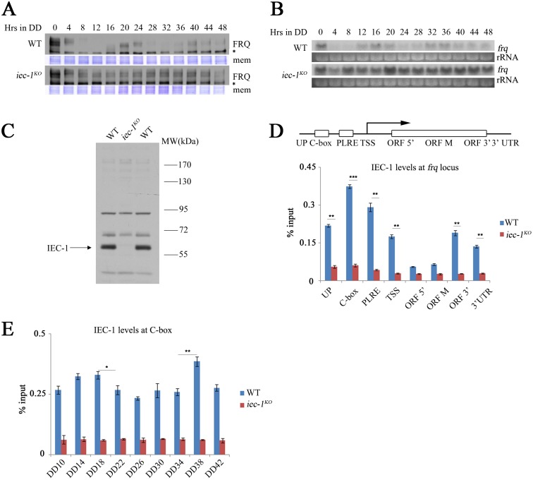
Fig 2. IEC-1 suppresses frq transcription and rhythmically binds to the frq promoter.
(A) Western blot analysis showing the circadian oscillation of FRQ proteins in the wild-type and iec-1KO strains. The strains were grown in 2% glucose liquid media. The asterisk indicates a nonspecific cross-reacted protein band recognized by our FRQ antiserum. The Coomassie Brilliant Blue-stained membranes (mem) represent the total protein in each sample and were used as a loading control. (B) Northern blot analysis of frq transcription in the wild-type and iec-1KO strains. rRNA was used as a loading control. The strains were grown in 2% glucose liquid media. (C) Immunodetection of IEC-1 protein in the wild-type strain and the iec-1KO mutant using antiserum that specifically recognizes the IEC-1 protein in the wild-type strain. The arrow notes the specific IEC-1 protein band detected by our IEC-1 antibody. The strains were grown in 2% glucose liquid media. (D) ChIP analysis showing the recruitment of IEC-1 at different regions of the frq locus in the wild-type and iec-1KO strains at DD18. The strains were grown in 2% glucose liquid media. C-box, clock box; PLRE, proximal light-regulated element; TSS, transcription start site; ORF, open reading frame; UTR, untranslated region. (E) ChIP analysis showing the enrichment of IEC-1 at the C-box of the frq promoter in the wild-type and iec-1KO strains at the indicated time points. The strains were grown in 2% glucose liquid media. Significance was assessed by two-tailed t-test. *P<0.05, **P<0.01. Error bars show the mean ±S.D. (n = 3).

