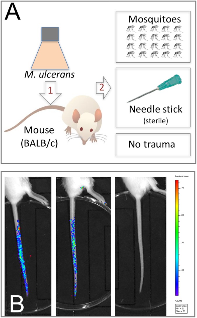
Fig 1. Schematic representations of the two BU transmission models tested in this study.
(A) Model-1 tests transmission of M. ulcerans present on a skin surface following a puncturing injury created by mosquito blood-feeding or needle stick. (B) Visualization of bioluminescent M. ulcerans JKD8049 (harbouring plasmid pMV306 hsp:luxG13) [29, 30] on the mouse-tail in model-1, showing the distribution of bacteria immediately after coating for two mice, versus an uncoated animal. M. ulcerans culture concentration used for tail coating was 8.3x10⁵ CFU/mL.

