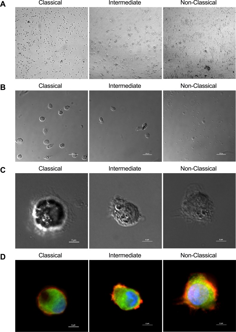
Fig 2. Morphology of sorted monocyte subsets.
(A) Low power magnification of monocyte subsets after magnetic enrichment and high-speed sorting from leukocyte concentrates (photomicrographs taken at 10X). (B) Differential interference contrast (DIC) image of monocyte subsets after two-day culture and adherence to substrate at low magnification (upper panel) and high magnification (lower panel). (C) Fluorescent image of the cells shown in lower panel B, with actin (orange), vinculin (green), β-tubulin (green) and nucleus (blue) stained to show morphological features.

