
Figure 2.
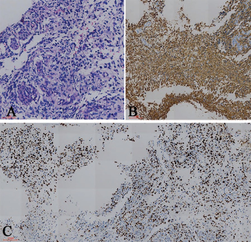
Histopathological and immunochemical findings. (A) Hematoxylin- and eosin-stained section revealed that the tumor is composed of cells with prominent eosinophilic cytoplasm, nuclear atypia, and microvascular proliferation. (B) Immunohistochemical staining shows positivity for glial fibrillary acidic protein. (C) The proliferation index (anti-Ki67) is 60% [magnification (A) 200×, (B), and (C) 100×].