
Figure 2.
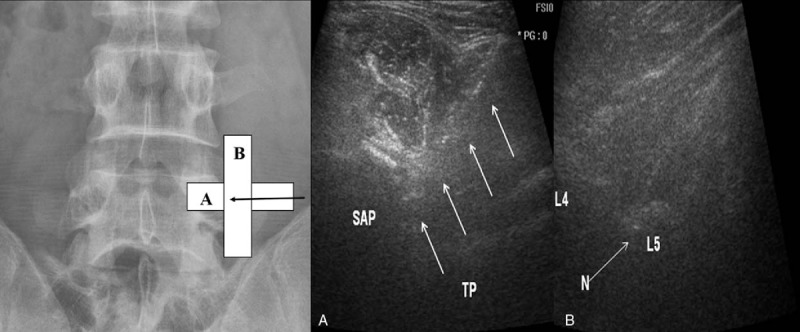
Ultrasound-guided medial branch block by a posterolateral approach with short-axis view and an in-plane free-hand technique. (A) The needle (arrow) is positioned using short axis in-plain approach to the angle between superior articular process (SAP) and the transverse process (TP) for a right-sided L4 medial branch block. (B) Check-up of the needle tip (N) positioned at the upper part of the L5 transverse process (L5) is facilitated using a long-axis and out-of-plane view.