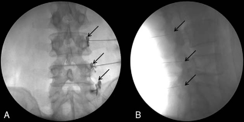
Figure 3.

Fluoroscopy-guided medial branch block. (A) Anteroposterior view. The contrast medium filled the L4/L5 superior articular processes for the L3/L4 medial branch block and the groove between the ala of the sacrum and the superior articular process of the sacrum for the L5 dorsal ramus. (B) Lateral view.
