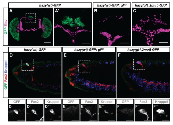
Figure 1.
Expression analysis of the hazy(wt)-GFP reporter in the ocelli and Bolwig's organ PRs. (A-C) In the case of the ocelli, these are 3 visual organs located dorsally on the head of adult flies (A). Samples were stained with antibodies against GFP (green), and Elav (used as a neuronal marker, magenta). The hazy(wt)-GFP reporter was expressed in the ocelli in wild-type (A, A'), but not glass mutant background (B). A hazy(gl1,2mut)-GFP reporter in which the 2 Glass binding sites were mutated was not expressed in the ocelli (C). (D-F) In the case of the Bolwig's organ, this is a larval eye that develops from the optic placode during embryogenesis. Stage 14 embryos were stained with antibodies against GFP (green), Fas2 (red) and Kruppel (blue). At this stage, the developing Bolwig's organ is located dorsally, still in contact with the surface, and can be identified both because of its position and the co-expression of Fas2 and Kruppel.,17,20,36 Similar to the ocelli, the hazy(wt)-GFP reporter was expressed in the Bolwig's organ in wild-type (D), but not glass mutant background (E). Also, hazy(gl1,2mut)-GFP was not expressed in the Bolwig's organ (F). For each image, the 3 channels from a close-up of the Bolwig's organ were separated and are shown below in grayscale (D′-F‴). Scale bars represent 20 μm in D′-F‴; 30 μm in A′, B-F; and 100 μm in A.

