
Figure 1.
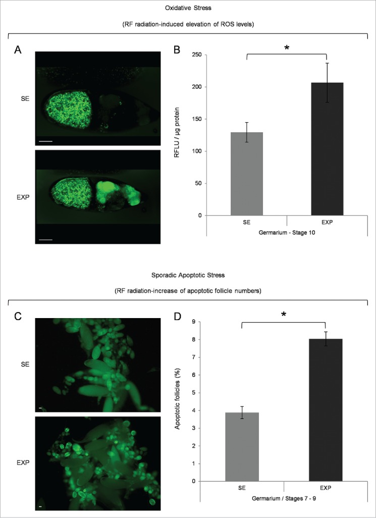
Mobile-phone radiation causes elevation of ROS cellular contents and activation of sporadic cell death, during early and middle oogenesis in Drosophila melanogaster. (A) Confocal laser scanning microscopy (CLSM) images illustrating representative ovarian follicles of stage 10, as they are stained with the general ROS-detector CM-H2DCFDA. Positively reagent-reacted nurse cells were detected only in ovaries of the exposed (EXP) flies (oocyte's green -spotty- signal emanates from auto-fluorescence of yolk granules). (B) Bar-graphs presenting ROS cellular levels ± SEM, as they are measured by suitably used fluorimetric assays, in control (SE: Sham Exposed) and truly exposed (EXP) follicles of all developmental stages from germarium (2b) up to stage 10. (C) Fluorescence microscopy images showing ovarian follicles from sham-exposed (SE) and truly exposed (EXP) flies, as they (follicles) are stained with the acridine orange apoptotic reagent. (D) The ratio of sporadically generated apoptotic follicles was estimated using the number of positively stained, with acridine orange, follicles, of early (germarium 2b) and middle (stages 7–9) oogenesis, against the total number of ovarian follicles of the same developmental stages ± SEM. Scale bars: 50 μm. N = 3, n = 3. SEM: Standard Error Mean. N: number of biological samples; n: number of biological experiments. *: P < 0.05.