
FIGURE 1. MAA causes extensive germ cell apoptosis but does not result in macrophage infiltration.
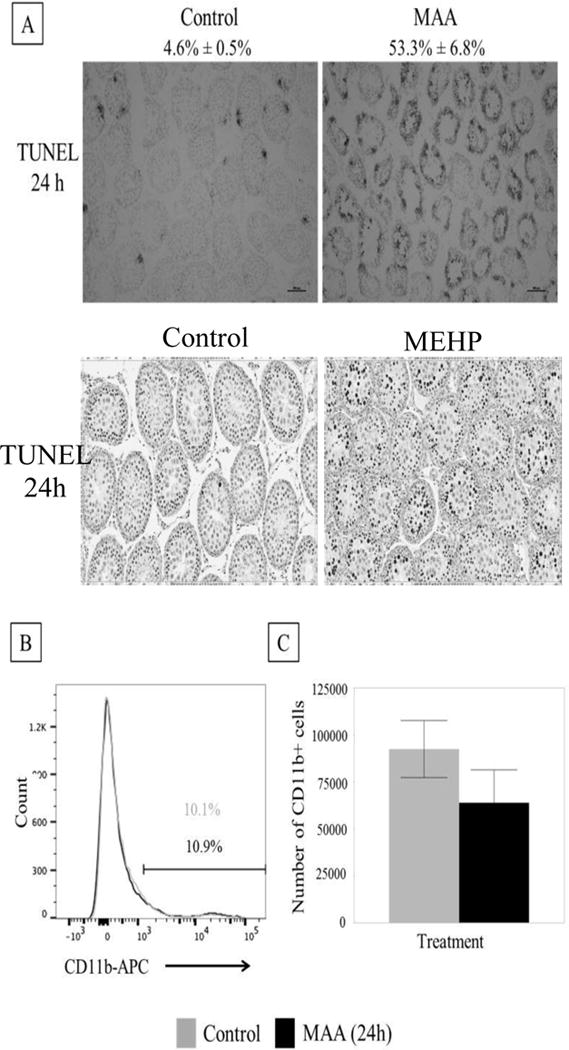
(A) Top: Representative photomicrographs of TUNEL staining on frozen MAA (650 mg/kg, p.o.) or equivalent volume control (5.5 ml/kg, NaCl) treated PND 28 rat testis after 24h exposure (n=3 per treatment). MAA induced a significant increase in apoptotic index (% seminiferous tubules with >3 TUNEL positive germ cells/total number of seminiferous tubules) from 4.6% ± 0.5% to 53.3% ± 6.8%. Bottom: Representative photomicrographs of TUNEL staining on paraffin embedded MEHP (667 mg/kg, p.o.) or equivalent control (2ml/kg corn oil) treated PND 28 rat testis after 24 h exposure. MEHP induced a significant increase in apoptotic index nearly 100 fold [9]. (B) Histogram of MAA or control treated rat interstitial cells (after 24h exposure). The CD11b+ population has high intensity fluorescence and is on the right of the graph. This population does not change between treatment (black bar) and control (grey bar). The population on the left is the CD11b- population. (C) When the percentage CD11b+ found from flow were applied to the total number of cells collected from the interstitium, there were no differences in the total number of CD11b+ cells. Statistical analysis was performed using Student’s t-tests.