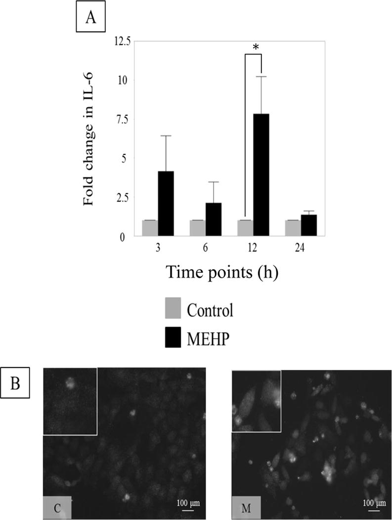
FIGURE 4. IL-6 increases in Sertoli cell rat line ASC-17D cells after in vitro exposure to MEHP.

(A) IL-6 expression in control (0.04% DMSO, control, grey) or MEHP (200 μM, black) treated ASC-17D cells at 3, 6, 12 and 24 h. MEHP induced a significant increase of IL-6 mRNA levels at 12 h. Statistical analysis was performed using two-way ANOVA for matched pairs with Sidak’s multiple comparison test and each MEHP-treated sample was compared to its time-point control. Representative microphotographs (B) of ASC-17D SC line immunostained with IL-6 (green, AlexaFlour488) after exposure to C (DMSO, control) or M (MEHP) for 24 h.
