
Figure 2. IL-6 expression is enriched in the stromal compartments of human PDAC tissue.
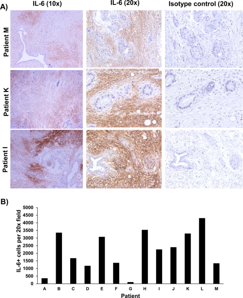
Human PDAC tumors (among n=13 patients) stained for IL-6 expression by immunohistochemistry. A) IL-6 (Brown Chromogen) IHC staining from three representative patient samples is displayed at 10× and 20× magnification. Tissue sections from each patient were stained with an isotype control Ab to account for background. B) Number of IL-6 positive cells per field was quantified.