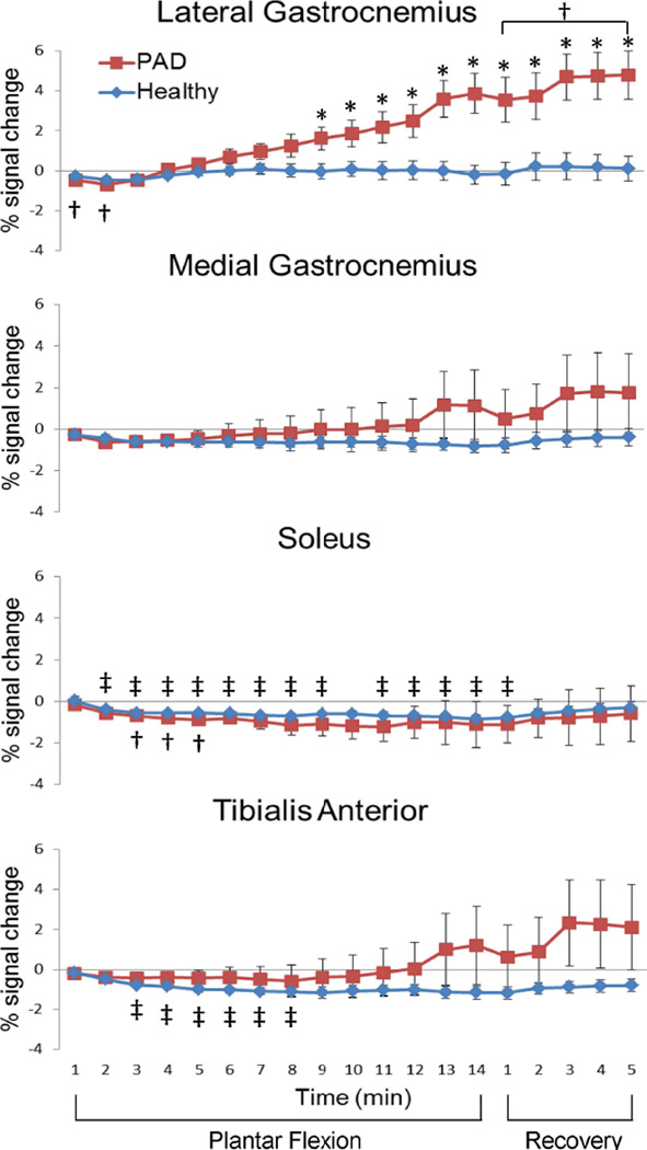
Figure 3.
T2*-weighted signal time courses of four calf muscles. % signal change comparing to the one-minute baseline (mean ± SEM) in a temporal resolution of one minute from healthy participants (n = 9, blue diamonds) and PAD patients (n = 8, red squares). †, significant signal change comparing to the baseline of the PAD participants, paired t-tests with Bonferroni correction, p < 0.05; ‡, significant signal change comparing to the baseline of the HC participants, paired t-tests with Bonferroni correction, p < 0.05; *, T2*-weighted signal in PAD patients significantly higher than that in HC participants, two-sample t-tests, p < 0.05.

