
Figure 3.
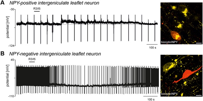
Activation of RXFP3 receptors excites or inhibits intergeniculate leaflet neurons, depending on their neurochemical nature (adapted from Blasiak et al., 2013). (A) A zero current‐clamp recording illustrating the depolarising effect of bath‐applied RXFP3 agonist, R3/I5 (100 nM, horizontal bar). Upwards deflections represent truncated action potentials present on top of calcium spikes evoked by membrane potential recovery from hyperpolarisation induced by current injection (downward deflections), and a confocal projection image of the neuron depolarised by R3/I5 stained for biocytin injected into the neuron (red) and neuropeptide Y (NPY) immunoreactivity (yellow) revealing the NPY nature of the neuron recorded. Scale bar, 10 μm. (B) A zero current‐clamp recording illustrating the hyperpolarising effect of bath‐applied R3/I5 (100 nM, horizontal bar) on the membrane potential and firing properties of another intergeniculate leaflet neuron, and a confocal projection image of the neuron hyperpolarised by R3/I5 stained for biocytin (red) and NPY immunoreactivity (yellow) revealing the NPY‐negative nature of the neuron recorded. Scale bar, 10 μm.