Figure 8.

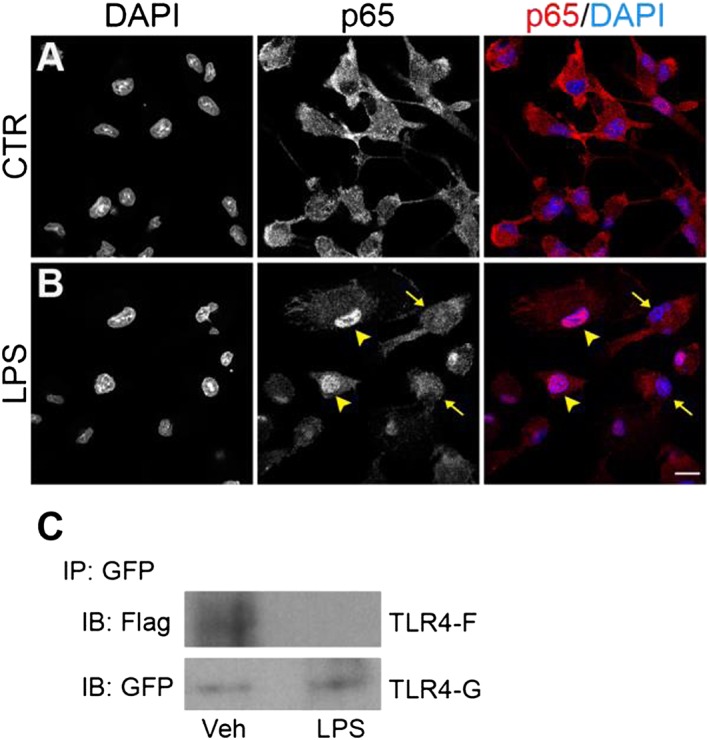
Effect of a low magnesium environment on NF‐κB p65 nuclear translocation and TLR4 dimerization. (A, B) Microglial cells, exposed to a low magnesium environment, were stimulated with 100 ng·mL−1 LPS for 90 min and further processed for NF‐κB p65 immunostaining. DAPI counterstaining is shown in the images in the left column and in the merged images in the right column. Experiments were performed at least five times, and representative confocal images showing subcellular localization of p65 in unstimulated and LPS‐stimulated microglia are shown in (A) and (B) respectively. Arrowheads point to examples of p65 nuclear localization, while arrows point to examples of cytoplasmic distribution. Scale bar, 10 μm. (C) Ba/F3 cells expressing TLR4‐Flag (TLR4F), TLR4‐GFP (TLR4G), MD2‐Flag and CD14, exposed to a low magnesium environment, were stimulated with 250 ng·mL−1 LPS for 30 min. Cell lysates were then immunoprecipitated with mouse anti‐GFP antibody and immunoblotted with mouse anti‐Flag (upper) or chicken anti‐GFP (lower) antibody. Experiments were performed five times, and representative results are shown. IB, immunoblot; IP, immunoprecipitation; Veh, vehicle.
