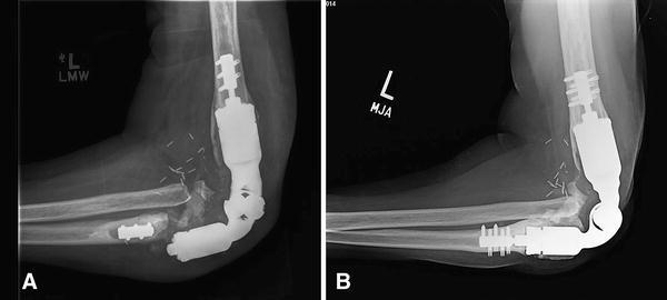
Fig 3A–B.

(A) A plain radiograph shows a well-fixed distal humeral compression implant adjacent to a failed ulnar compression component in Patient 2. The mode of failure was lack of ingrowth, periprosthetic fracture at the bone-prosthesis interface, and prosthetic fracture through the titanium traction bar. (B) The patient underwent revision surgery with implantation of another compression endoprosthesis.
