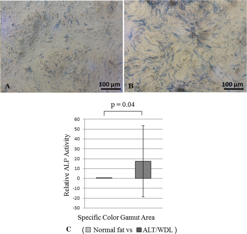
Fig. 3A–C.
The photomicrographs show MSCs derived from (A) normal fat after 14 days of induced osteogenic differentiation, and from (B) ALT/WDL after 14 days of induced osteogenic differentiation. ALP staining was performed in both cases. Greater ALP staining was observed in MSCs derived from ALT/WDL. (C) A graph shows the osteogenic differentiation ability of MSCs derived from normal fat and ALT/WDL using ALP staining. Osteogenic differentiation potency was assessed by measuring a specific colored area using Image J image analysis software. MSCs derived from ALT/WDL had higher differentiation potency than MSCs derived from normal adipose tissue. ALP = alkaline phosphatase.

