Figure 1.

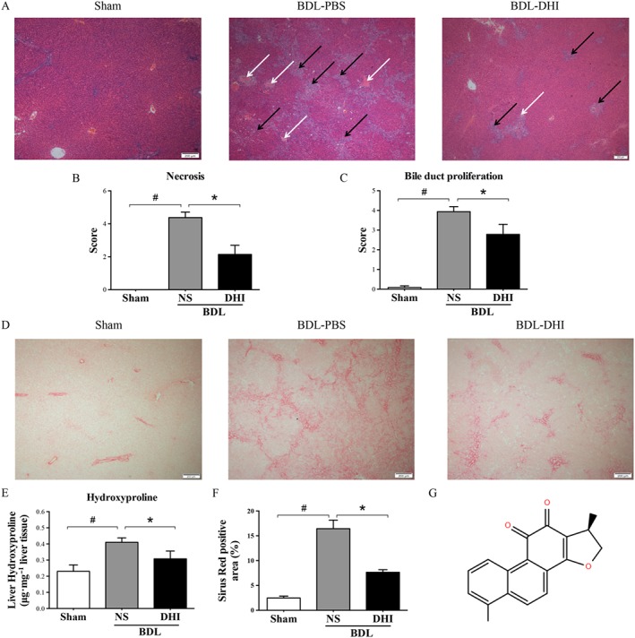
(A) Liver pathological changes were detected by H&E staining (magnification × 40). The necrosis and bile duct proliferation areas have been indicated by white and black arrows respectively. Double‐blinded quantitative assessment of (B) hepatic necrosis and (C) liver bile duct proliferation showed that DHI ameliorated BDL‐induced histopathological changes. (D) The degree of liver collagen accumulation was determined by Sirius red straining (magnification × 40). (E) Percentage of Sirius red positively strained areas and (F) liver hydroxyproline concentration assays demonstrated that DHI reduced liver fibrosis in BDL rats. (G) The structure of DHI. The values are expressed as the mean ± SEM (n = 6 in sham group; n = 7 in BDLNS/BDL‐DHI group), #P < 0.05; significantly different from sham group, * P < 0.05; significantly different from BDL‐NS group; ANOVA followed by Tukey's test.
