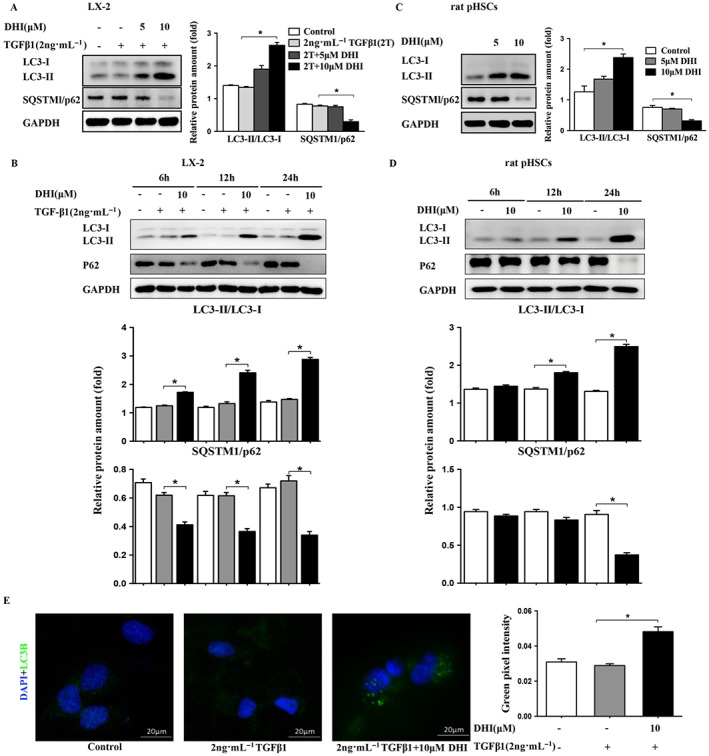
Figure 6.

DHI enhanced autophagy in HSCs. DHI dose‐dependently and time‐dependently enhanced the conversion of LC3‐I to LC3‐II and reduced the protein expressions of SQSTMl/p62 in (A and B) LX‐2 cells and (C and D) rat pHSCs. (E) LX2 cells were treated with DHI, and LC3 puncta formation was analysed by immunofluorescence; original magnification ×400. Quantitative analysis of green pixel intensity was also performed by image J. The data are expressed as the mean ± SEM of five independent assays, #P < 0.05; significantly different from the control group and *P < 0.05; significantly different from the TGFβ1 treatment group in LX‐2 cells; *P < 0.05; significantly different from the control group of rat pHSCs; ANOVA followed by Tukey's test.
