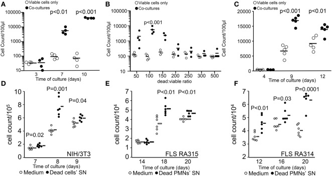
Figure 1.
Stimulation of tumor cell proliferation by bystander dying cells, the “feeder cells” effect. The upper panel shows viable cell counts of cocultures [closed circles in panels (A–C)] and viable cells alone in D10 medium [open circles in panels (A–C)]. Cocultures were composed of viable (200 cells/well) and lethally ultraviolet light type B-irradiated (10,000 cells/well) B16F10 melanoma cells (A). Dead/viable ratio titration of cocultures (B). Transwell cocultures with 10,000 apoptotic cells (C). The lower panel shows cell counts of independent wells containing fibroblasts in R10 medium [open circles in panels (D–F)] or in supernatants (SN) from apoptotic cells [closed circles in panels (D–F)]. Mouse NIH/3T3 fibroblast (1,000 cells/well) cultures with SNs of 0.2 million cells/ml apoptotic homologous cells (D). Human fibroblast-like synoviocytes (FLS, 1,000 cells/well) from two rheumatoid arthritis patients RA315 (E) and RA314 (F) cocultured with SNs from apoptotic neutrophils (0.2 million cells/ml).

