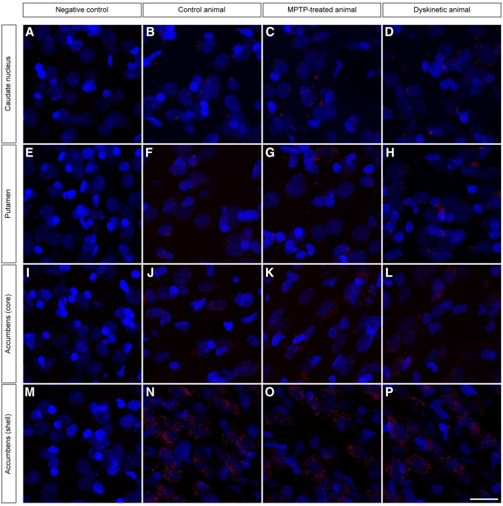
Fig. 4.
PLA-based detection of D1–D2 receptor heteromers. The use of the PLA protocol enabled the unequivocal detection of D1–D2 receptor heteromers in all striatal territories (caudate nucleus, putamen and the core and shell subdivisions of the accumbens) across all animal groups (control, MPTP-treated and dyskinetic animals). Negative control experiments, conducted the same way by avoiding the conjugation of the anti-D2 antibody with the Duolink Probemaker kit, resulted in a complete lack of stain. Each D1–D2 receptor heteromer is visualized as a single red spot. Cell nuclei are counterstained with Topro-3 (blue color). The strongest PLA stain is typically observed in the shell of the nucleus accumbens, followed by a gradual decline when considering the core of the accumbens, the putamen and the caudate nucleus. This pattern of staining is maintained in all the different animal groups. Scale bar 20 mm in all panels

