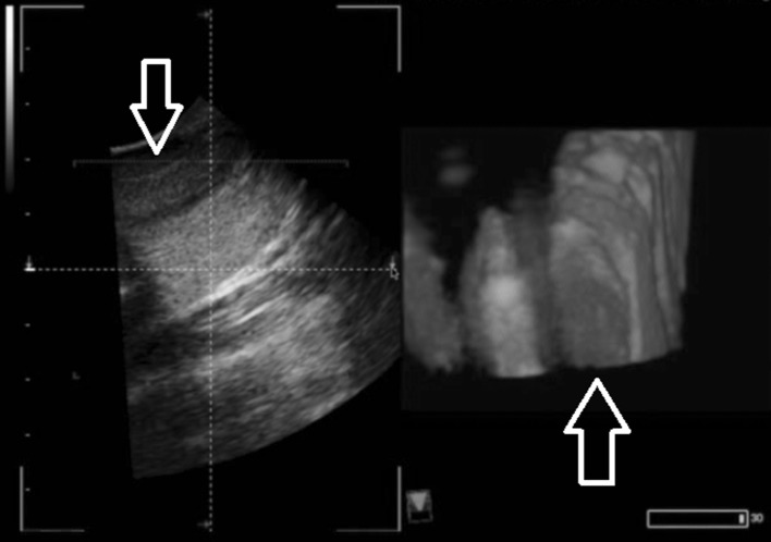
Fig. 5.
Incidental nodule found during ultrasound training. 2D ultrasound was used initially, as the thyroid nodule was discovered (left). Imaging using 3D/4D ultrasound (right) depicts the nodule and allows to see the nodule with both height and width from this viewpoint. The 3D/4D image can be rotated to further investigate the depth of the nodule, allowing for a better understanding of the size and shape of the nodule

