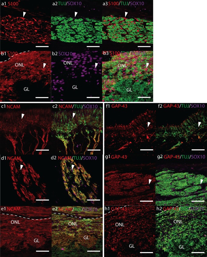
Fig. 1.
Confocal fluorescent micrographs showing sagittal cryosections of 17 pcw human foetal olfactory system immunolabelled with antibodies towards a, b TUJ (green), S100 (red) and SOX10 (magenta), c–e NCAM (red), TUJ (green), SOX10 (magenta) and f–h GAP-43 (red), TUJ (green), SOX10 (magenta). Scale bar 40 μm, dashed white lines indicate the surface of the olfactory bulb. a Precise cross-section of an olfactory nerve showing numerous SOX10/S100+ OECs (white arrowheads) surrounding bundles of TUJ+ olfactory axons. b SOX10/S100+ OECs (white arrowheads) are observed throughout the olfactory nerve layer (ONL) and surrounding but rarely within the glomerular layer (GL). c, f Olfactory receptor neurons (white arrowheads) and their axons co-labelled with TUJ and NCAM or GAP-43, respectively. d, g Cross-section of an olfactory nerve showing co-labelling of TUJ+ olfactory axons with NCAM and GAP-43. Co-labelling with SOX10 is not observed (white arrows). e, h Co-labelling of the ONL and GL with TUJ and NCAM or GAP-43, respectively

