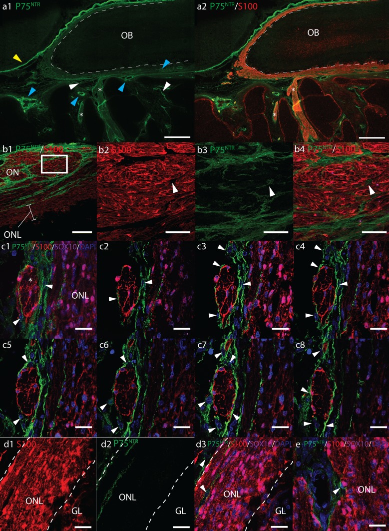
Fig. 3.
Fluorescent micrographs of sagittal cryosections from 17 pcw human foetal olfactory system immunolabelled with antibodies towards P75NTR (green), S100 (red), SOX10 (magenta), and DAPI nuclear stain (blue). a Axioscan micrograph montages of the olfactory bulb (OB) and olfactory nerves (ON, asterisk). Scale bar 500 μm. S100 labels the entire olfactory nerve layer (ONL) and ONs, whereas P75NTR surrounds the outside of the OB and ONs. P75NTR labelling was also observed surrounding blood vessels (blue arrowhead); on cells within the nasal stroma (white arrowheads) and intensely expressed in meningeal tissue (yellow arrowhead). b Cross-section of olfactory nerves showing P75NTR+ processes (white arrows) surrounding S100+ OECs. b1 Scale bar 100 μm. b2–4 Scale bar 20 μm, shows magnified images of area within white box in b1. c2–7 Serial optical sections through a 13 μm z-stack (c1) showing DAPI labelled nuclei and P75NTR+ processes of perineurial cells (white arrows) surrounding S100+ OECs within a cross-section of an olfactory nerve (asterisk). Scale bar 20 μm. d1–3 In the outer ONL, P75NTR is expressed on cells (white arrowheads) surrounding ONs as they enter the ONL, but not on SOX10/S100+ OECs. Dashed white lines show rough boundary of ONL, scale bar 20 μm. e P75NTR+ cells surround presumptive arterioles in the ONL, scale bar 20 μm

