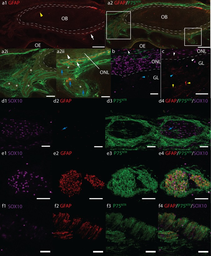
Fig. 4.
Axioscan fluorescent montages (a) and confocal fluorescent micrographs (b–f) of GFAP (red), P75NTR (green) and SOX10 (magenta) immunoreactivity in sagittal cryosections of a–d 17 pcw and f 12 pcw human foetal olfactory system. Scale bars a1, a2 500 μm, a2i 200 μm, a2ii 100 μm, b–e 40 μm. f 20 μm. Dashed white lines roughly denote inner and outer boundaries of the olfactory nerve layer ONL, olfactory bulb OB, olfactory epithelium OE, glomerular layer GL, olfactory nerves ON asterisk. Boxes in a2 show position of magnified areas in a2i and a2ii. a GFAP immunoreactivity in the ONL and ONs is sparse. Yellow arrowhead a1 shows GFAP+ astrocytes, a2i shows the majority of ONs asterisks do not express significant GFAP, white arrowhead a2ii shows GFAP+ cells in ONL, blue arrows show sporadic GFAP in olfactory nerves asterisk. b, c GFAP in the ONL and GL, white and blue arrowheads show GFAP+ cells in ONL and glomerular layer, respectively. Yellow arrowheads show GFAP+ presumptive astrocytes in the deeper layers of the OB. d Sporadic GFAP in olfactory nerves which are surrounded by P75NTR+ perineurial cells. Co-labelling of GFAP & P75NTR is not observed. Pattern of GFAP and P75NTR co-labelling in peripheral facial nerves e is similar to that seen in occasional small nerve bundles in nasal stroma f and a2ii white arrow

