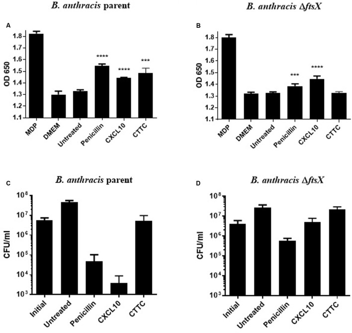
FIGURE 5.
Detection of peptidoglycan release using HEK cells that express a NOD2-NF-κB reporter system with concurrent CFU data collection. Peptidoglycan subunit release into the supernatant of untreated or antimicrobial-treated vegetative cells was detected by an HEK-BlueTM hNOD2 mammalian cell line that expresses the NOD2 receptor, which detects bacterial peptidoglycan and activates an NF-κB reporter system that produces a SEAP enzyme for a colorimetric-based quantitative readout. (A) Detection of B. anthracis parent strain peptidoglycan release in response to penicillin (0.5 μg/ml), CXCL10 (1.4 μM), or CTTC (2.8 μM), as compared to the untreated control group of the same bacterial strain. (B) Detection of B. anthracis ΔftsX peptidoglycan release in response to penicillin (0.5 μg/ml), CXCL10 (1.4 μM), or CTTC (2.8 μM), as compared to the untreated control group of the same bacterial strain. CFU enumeration was performed at the 2 h time point in parallel with supernatant collection and peptidoglycan release assay. These corresponding CFU data are shown for (C) B. anthracis parent strain or (D) B. anthracis ΔftsX exposed to penicillin (0.5 μg/ml), CXCL10 (1.4 μM), or CTTC (2.8 μM). Results are represented ± SEM, n = 3 separate experiments with triplicate wells used in each experiment. ∗∗∗p ≤ 0.0001, ∗∗∗∗p < 0.0001.

