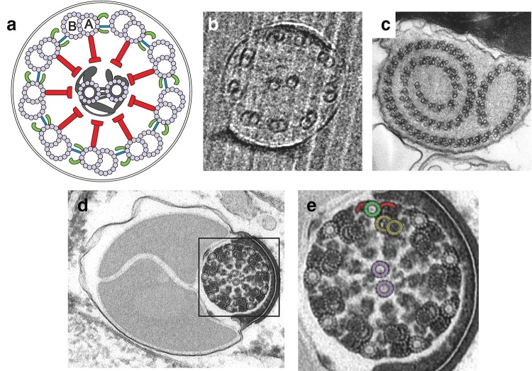
Fig. 2.
Exemplary axonemal structures found in sperm. a schematic diagram of a cross-section from a canonical 9 + 2 axonemal structure. View from the head towards the flagellar tip. Nine microtubule doublets (with subtubules A and B) are arranged cylindrically around an additional pair of microtubule singlets located at the centre. Two dynein arms (green) containing different subsets of dynein motors are attached to subtubule A and point towards the next B-tubule of the neighbour microtubule doublet in a clockwise direction (green). Dynein motors, fuelled by ATP, move adjacent microtubule doublets along the axis of the axoneme. Microtubule sliding is thought to be transformed into bending by mechanical constraints produced by their attachment to the basal body, nexin links (blue) (Lin et al. 2012), or clustering of motor activity (Movassagh et al. 2010). Radial spokes (red) connect the nine doublets in the periphery with the inner microtubule pair and associated filaments (grey). This complex is thought to orchestrate microtubule activity (Wargo and Smith 2003). b Flagellar cross-section from the sea urchin Arbacia punctulata with the canonical 9 + 2 structure. c Cross-section of the aberrant axoneme from the fly Sciara coprophila displaying its spiral shape. The axoneme is composed of 60–90 doublets, each one associated with a singlet or accessory microtubule. Subtubule A has two dynein arms (Jamieson et al. 1999). d Flagellar cross-section from the fly Drosophila bifurca. The axoneme and the two large mitochondrial derivatives can be seen. e Magnified detail from panel d displaying the characteristic 9 + 9 + 2 axoneme from insects. An accessory tubule (green) with its corresponding arms (red), a microtubule doublet (yellow), and the central pair (violet) have been labelled for clarity. Images are courtesy of Drs. S. Irsen (b) and R. Dallai (c–e)

