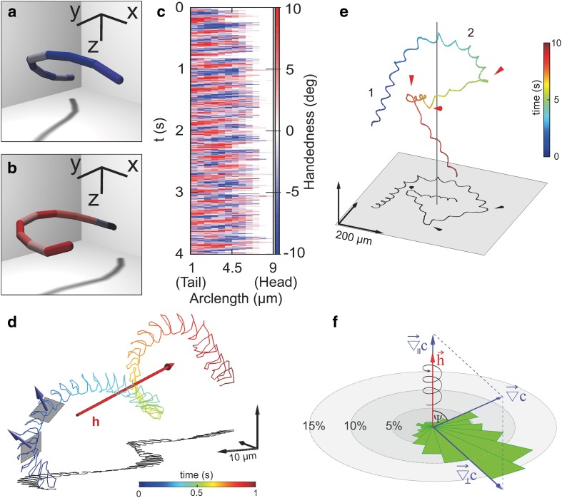
Fig. 3.
Recent advances in 3D sperm motility. a–c 3D reconstruction of the flagellum from malaria sperm. Flagella display a left (a) or right (b) handedness. The spatiotemporal map of chirality (c) shows propagating waves of alternate handedness across the flagellum. d 3D swimming path of a freely moving sperm cell from the sea urchin A. punctulata. Sperm swim along a helical path. From the motion of the sperm head, the flagellar beat plane can be inferred. Helix axis is shown in red. Two best-fitting planes along the path and the direction normal to them are shown in grey and blue, respectively. Time is color-coded along the path (see corresponding colorbar). e Swimming path of a sperm cell navigating in a chemical gradient. The axis of symmetry of the gradient is indicated by a grey line at the center. The cell approaches the gradient following a regular helical path (1). The helical axis bends smoothly during chemotaxis (2). Smooth helix alignment is interrupted by abrupt steering events (indicated by red cones). Strong alignment events occur when smooth alignment does not suffice to keep up-gradient swimming. f Rose plot of alignment events showing the changes in direction of the helical axis with respect to the parallel () and perpendicular () gradient components. Alignment of the helical axis is not random but scatters around the perpendicular gradient component, showing that the helical axis bends deterministically to align with the gradient direction

