Figure 2.

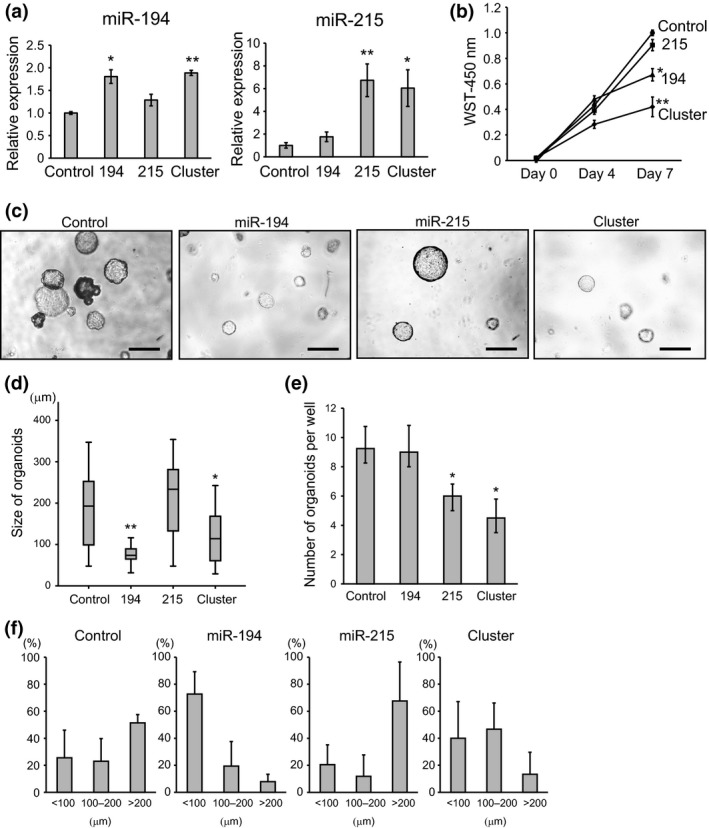
Growth and morphological change in intestinal tumor organoids transfected with miR‐194 and miR‐215. (a) Relative expression levels of miR‐194 and miR‐215 in miRNA‐transfected intestinal tumor organoids. U6 was used as an internal control. (b) WST cell proliferation assay of intestinal tumor organoids transfected with miRNA. (c) Representative images of intestinal tumor organoids transfected with miR‐194, miR‐215 and cluster. Scale bars: 1000 μm. (d) Size of intestinal tumor organoids transfected with miR‐194, miR‐215 and cluster. (e) Number of intestinal tumor organoids transfected with miR‐194, miR‐215 and cluster. (f) Size distribution of organoids normalized to the total number of organoids for each treatment. *P < 0.05, **P < 0.01. Control, 194, 215 and cluster represent tumor organoids transfected with empty vector, miR‐194, miR‐215 and cluster, including miR‐194 and miR‐215, respectively.
