
Figure 1.
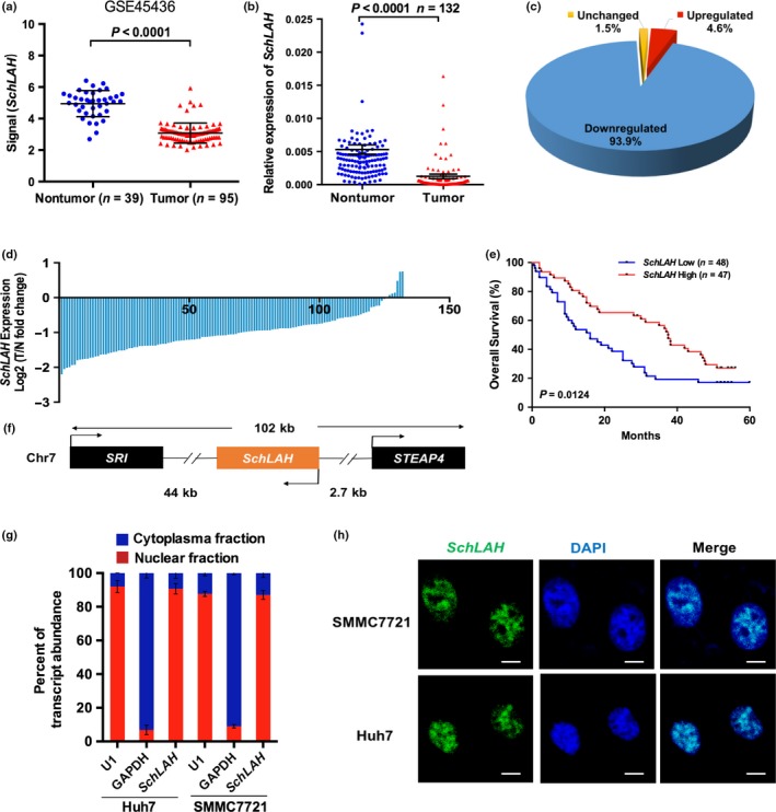
Identification of SchLAH as a HCC‐associated lncRNA. (a) SchLAH was downregulated in tumor tissues compared to nontumor liver tissues (GSE45436). (b–d) SchLAH was downregulated in tumor tissues compared to paired adjacent noncancerous liver tissues (n = 132). The expression level of SchLAH was analyzed by RT‐PCR and normalized to β‐actin. (e) Survival rates of 95 HCC patients who underwent liver surgery were compared between the SchLAH high‐expression and SchLAH low‐expression groups (log rank: P = 0.0124). The median expression level was used as the cutoff. (f) Schematic representation of SchLAH location on chromosome. (g) Fractionation of Huh7 and SMMC7721 cell lysates demonstrated nuclear expression of SchLAH. U1 RNA served as an internal control for nuclear gene expression. Values are mean ± standard deviation (n = 3). (h) Nuclear localization of SchLAH detected by RNA FISH in SMMC7721 and Huh7 cells. Scale bar, 10 μm.