Figure 3.

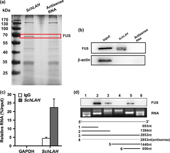
The Interaction of SchLAH with FUS. (a) SchLAH and FUS specifically interacted in vitro. SDS‐PAGE gel of proteins bound to SchLAH (left lane) or antisense RNA (right lane). The highlighted region was submitted for mass spectrometry identifying FUS as the band unique to SchLAH. (b) Western blot analysis of the specific association of FUS with SchLAH. A nonspecific protein (β‐actin) is shown as a control (n = 3). (c) RNA immunoprecipitation (RIP) experiments were performed using the FUS antibody, and specific primers were used to detect SchLAH or GAPDH. Values are mean ± standard deviation (n = 3). (d) SchLAH bound to FUS/TLS through a region between 800 and 1800 nt which contains “GU” rich motif.
