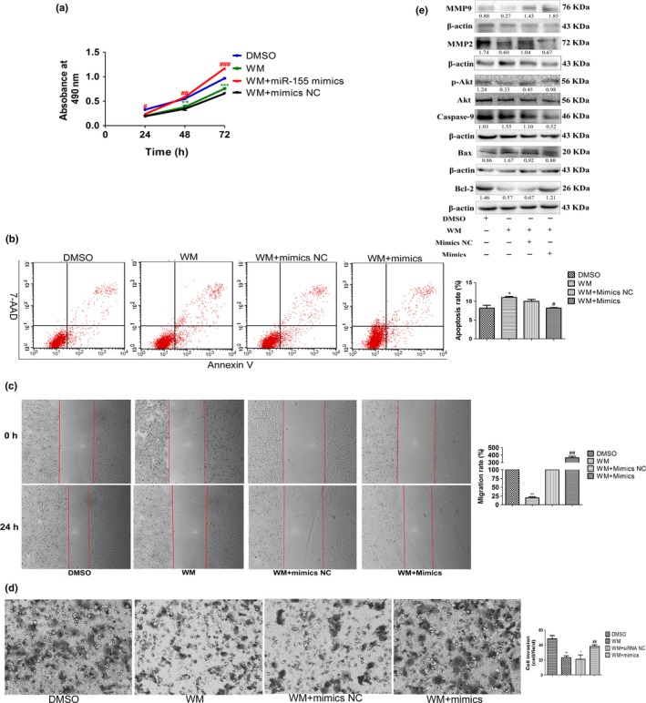
Figure 5.

MiR‐155‐5p promotes proliferation, invasion and migration, and inhibits apoptosis by the PI3K/Akt pathway. (a–e) Cell viability, the percentage of apoptotic cells, wound healing and transwell assay, and the expression of Akt and p‐Akt, Bcl‐2, Bax, caspase‐9, MMP2 and MMP9 in Hep3B cells treated with DMSO, wortmannin alone, wortmannin and miR mimics NC, and wortmannin and miR‐155‐5p mimics; the intensity of each band was quantified; the value under each lane indicates the relative expression level of the regulators; n = three repeats with similar results, *P < 0.05, **P < 0.01, versus Hep3B treated with DMSO; #P < 0.05, ##P < 0.01, ###P < 0.001, versus Hep3B treated with wortmannin alone by Student's t‐test. WM, wortmannin.
